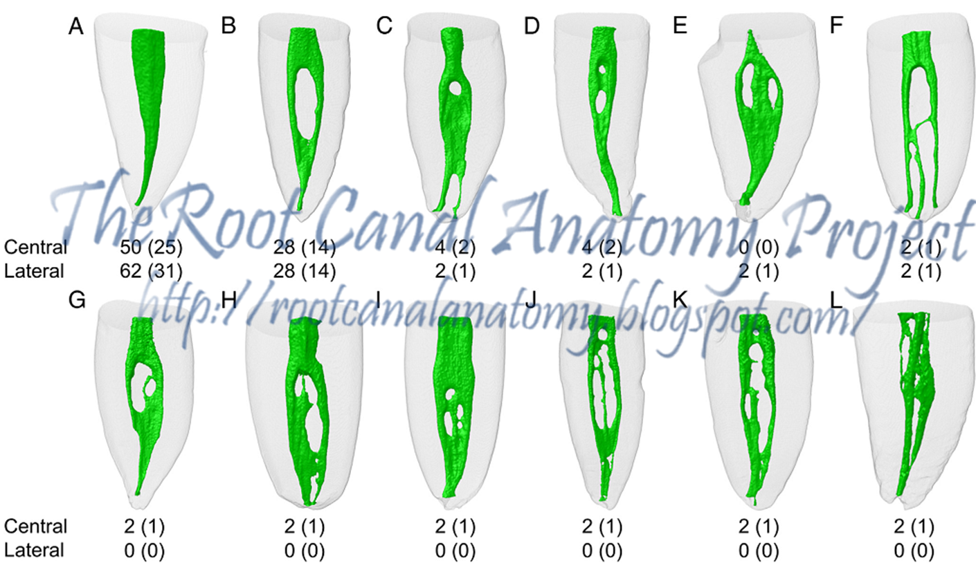
#Lower macillary central invisor canal crack#
Their magnitude is important because crack initiation and propagation are mainly controlled by tensile strain. īecause a relationship between lateral compaction and root fracture has been proposed, the characterization of strains created in radicular dentin during lateral compaction has been a research area of much interest. Such micro-cracks in radicular dentin might be created by rotary NiTi instruments through the process of canal instrumentation. These cracks may subsequently develop into vertical root fracture (VRF). Such stresses are a concern, especially when preexisting micro-cracks are present in the dentin, with potential propagation from a subcritical to a critical length. The apical force applied through finger spreaders during compaction of gutta-percha, which is currently the most commonly used root filling material, exerts pressure on the material, resulting in circumferential tensile stresses on the canal surface. They generate less stress during lateral compaction, have better penetration capabilities and result in decreased apical microleakage in endodontically treated teeth. Nickel-titanium (NiTi) finger spreaders are preferred over the less elastic stainless steel ones. Such spreaders exert less lateral force on the canal walls and allow for greater tactile sense. Traditionally, lateral compaction was applied using hand spreaders, but it is currently applied mainly using finger spreaders. Other maxillary lateral incisors show twisted roots or distorted crowns.Lateral compaction, frequently cited as the 'gold standard' technique for root canal filling, is one of the most commonly used methods and is utilized by more than half of all practitioners worldwide. One type of malformed maxillary lateral incisor displays a large, pointed tubercle as part of the cingulum some have deep developmental grooves that extend down the root lingually with a deep fold in the cingulum. Maxillary lateral incisors are more likely to be congenitally missing than any other teeth except the third molars. For some, the lateral incisors are missing entirely. A common situation is to find maxillary lateral incisors that have a nondescript, pointed form such teeth are called peg-shaped lateral incisors. If the variation is too great, it is considered a developmental anomaly. Maxillary lateral incisors vary in form more than any other teeth in the mouth except the third molars. These teeth differ from the central incisors in that their individual development varies considerably. The features-curvatures, concavities, and convexities-of the lateral incisors are more prominent and show more distinction and contrast than those of the central incisors. Lateral incisors are small in all dimensions except root length. Maxillary lateral incisors complement the central incisors in function and resemble each other in form. The root is usually 2 to 3 mm longer than the crown, although the root-to-crown ratio varies considerably. The root of a central incisor from the labial aspect is cone shaped and has a blunt apex in most instances. However, normal use eventually wears down the rounded ridge into a flat edge, and therefore the term incisal edge is more appropriate than ridge. This ridge portion is then called the incisal ridge. When an incisor first erupts, the incisal portion of the crown is rounded and the mamelons are quite distinct. The incisal outline is usually regular and straight across the incisal ridge after the tooth has been in function long enough to wear down the mamelons. The cervical line crests slightly distal to the center of the tooth.

( Modified from Zeisz RC, Nuckolls J: Dental anatomy, St Louis, 1949, Mosby.)įrom this facial view, the distal outline of the crown is more rounded or convex than the mesial outline, the height of curvature being higher toward the cervical line. A, Labial view B, lingual view C, incisal view D, mesial view E, distal view.


 0 kommentar(er)
